This and That: Ehrenberg’s Diatoms

1a Diatoms

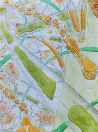
Images from E. César’s Tweet on the Ehrenberg Collection at the Museum für Naturkunde in Berlin.

Though I have more time to think deeply right now than ever in my life, I’m finding it difficult to do; everything is so different from usual that it’s unsettling.  That’s why I’m not focusing on one topic for a month’s worth of posts as I usually do, but flitting from one topic to another from week to week.  In part this is because of Twitter, my lifeline to the botanical world at the moment.  Thank goodness botanists are interesting people and post interesting ideas.  Most days I find at least one item worth bookmarking and then delving into more deeply.  That’s how I discovered Christian Gottfried Ehrenberg (1795-1876).  I must have come across his name in the past, especially when I was reading about Alexander von Humboldt because Ehrenberg accompanied the explorer on his trip to Siberia in 1829.

A Tweet on Ehrenberg by Edgley César, curator of diatoms at the Natural History Museum, London, included the image above.  It was the photo on the upper right that first caught my eye—obviously old data—and the illustration on the lower left was another lure.  César took the pictures at the Museum für Naturkunde in Berlin where he had spent a week examining specimens of a genus Ehrenberg had described and was amazed by how much work this “founding father” of diatom research had done and how well he drew.  As the thread continued, someone asked about Ehrenberg and César pointed them, and me, to a series of papers published in 1998 dealing with his life, work, and collections.

Ehrenberg was definitely productive throughout his life.  Born near Leipzig, he attended the university there, completing his doctorate on fungi in 1818.  His fungal herbarium is in the Botanic Garden and Botanical Museum Berlin-Dahlem.  From 1820-1825, Ehrenberg participated in an expedition to the Middle East, during which he and his friend Wilhelm Hemprich amassed 114 boxes with 46,000 plant and 34,000 animals specimens as well as seeds, fossils, minerals, and of course, mummies.  Yet the trip was grueling, with three-quarters of team dying, including Hemprich.  Ehrenberg published, Symbolae Physicae, a multivolume work on all aspects of the collection and including 800 plates, many based on his drawings.  He did not describe many of the plants he collected and left the world of higher plants to concentrate on microscopic work, on what were called infusoria, organisms found in decaying matter.  However, he did teach all his children to press plants and create their own herbaria.

A great deal of Ehrenberg’s research was on radiolaria and diatoms.  He considered them all tiny animals and carefully studied their internal structures, which he interpreted as digestive, reproductive, and muscular.  He thought that when better microscopes were developed, these organelles would be seen more clearly.  It is interesting that when diatoms were finally recognized to be more closely related to plants than animals, interest in their internal structures waned, and their taxonomy became based primarily on their elaborate silicate shells that come in a dizzying array of patterns.  The assumption became that there was little difference among these organisms internally; plant cell structures were just not that interesting.  Ancient shells found in diatomaceous earth have long been used in geological exploration, since they are related to oil deposits, but even present-day species are often dried, and just their shells examined.

Ehrenberg made extremely detailed and exquisite illustrations of these organisms and in 1838 published a book with 64 plates on Infusoria in all of their complexity.  He also kept detailed notes on his work, as well as retaining the specimens he’d examined.  Glass slides and coverslips were expensive, so he used small mica discs with a bit of Canadian balsam, a shorthand term for a thick liquid made from the tree’s resin that was a mainstay for 19th-century microscopists because of its optical properties.  Ehrenberg highlighted interesting organisms with small circles, and then with a little more balsam, stuck the discs to his notes.  These have been preserved for almost 200 years, though not without difficulties.

The Ehrenberg Collection at the Museum für Naturkunde consists of 40,000 microscope preparations, 5,000 raw samples, 3000 illustrations, and 800 letters.  It is the combination of different kinds of information that makes it so impressive and valuable, but also daunting.  Most of Ehrenberg’s vascular plant herbarium was at the Berlin-Dahlem botanic garden and was lost when its herbarium was bombed during World War II.  The infusoria, on the other hand, were at the museum and survived but in what would become East Berlin.  The collection was not curated or organized until after German reunification when new resources became available.  It was in light of this that the 1998 article collection was published to showcase Ehrenberg’s work and how the collection could be used, just as César is now using it.  The notes are now beautifully curated (see below), but this required a great deal of work.  The balsam has become brittle, and the mica discs are fragile and difficult to handle.  Over the years some had become unstuck, shifted, and were crushed.  Conservation was necessary because the records contain many type specimens, though as David Mann notes in the last article in the collection, types can present difficulties in terms of hunting them down in a compilation this vast and with all the vagaries it has been through.

1 Conserved

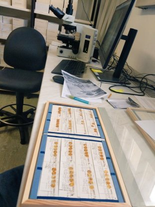
Photo of portion of conserved Ehrenberg Collection at the Museum für Naturkunde in Berlin.

As someone who is fascinated by diatoms, the Ehrenberg Collection is definitely a treasure (see video), along with the diatom collections at the Academy of Natural Sciences herbarium in Philadelphia (see earlier post) and at the Natural History Museum, London.  If you are interested in these beautiful organisms that are classified as algae, you might want to look at Martyn Kelley’s long-running Microscopes and Monsters blog where he deals with microscopic algae and environmental monitoring.

The Algal World: More Diatoms

kelly

Round Loch of Glenhead, watercolor by Martyn Kelly.

I have a bit more to add on diatoms; there just wasn’t enough space in my last post. I have already mentioned diatomaceous earth, composed of the remains of ancient diatoms. It is used for polishing because of the abrasiveness of the glassy shells made primarily of silica. The tiny, hollow shells have a great deal of surface area making them effective filtering agents as well. The earth can also be employed as an insecticide because the abrasiveness damages the waxy covering on many insects. Perhaps the greatest impact of diatoms on our life style today is that their internal remains are a major component of fuel oil, which was created from deposits of many kinds of plant material including the aquatic variety. In fact, the diatom contribution was so significant that some researchers today are considering diatoms as a form of future biofuel, in part because, among phytoplankton, a higher proportion of their fatty acids are monounsaturated making them a better starting material for biofuel.

One of the best and most engaging ways to learn more about diatoms and about how they are used to evaluate aquatic environments is to read the posts on Martyn Kelly’s Of Microscopes and Monsters blog. Kelly heads a consultancy firm in Britain that specializes in studying freshwater ecosystems, and diatoms are key to this research. He is an expert in the field but he writes very appealingly for nonspecialists. He has even produced a book on the subject that is available for a free download from his website. He describes what a diatom population tells about water quality, and how this changes with season and location. Kelly has been doing this work long enough that he can also discuss how things have changed—for better or worse—over time. He travels widely on business so there are posts on his experiences studying diatoms and other phytoplankton in many locations; there have been recent posts from India.

I should note that Kelly can definitely be opinionated in his posts, which is not a bad thing. He provides a sense of the political and economic issues surrounding diatoms—they are not only about beauty. As an independent consultant, he is a little freer to speak his mind, and as a Briton facing exit from the European Union, he has some interesting things to say that what this might or might not mean for environmental regulations in the United Kingdom. Kelly also looks at the local level, at such issues as how a new housing development is likely to affect nearby lakes and streams and what can be done to mitigate the changes. So there are many reasons why his writing is worth checking out.

What makes Kelly’s website particularly interesting is that he is also an artist, who returned to school to take up art seriously. I don’t think it’s a coincidence that he chose to focus on diatoms, because as I said in my first post on algae, they are particularly appealing aesthetically, and this is especially true of diatoms. In his effort to communicate about aquatic environments, Kelly sometimes includes his watercolors in posts. They are beautiful and portray the atmosphere of an underwater world. He includes not only diatoms but other planktonic organisms that would be associated with them in a particular body of water. Some of these works are also included in his book. I think it’s particularly powerful when a biologist is also an artist, because they bring knowledge and years of observation to their art. Kelly presents diatoms in a very different way than does Haeckel, whose portrayals are greatly enlarged, with all the structural details laid out for the viewer as static, structural monuments. Kelly’s diatoms, on the other hand, do not have as many details, but they are shown as immersed in their environment rather than separate from it, and there is a sense of movement. I see these two approaches as complementary and definitely worth studying. We are very fortunate that both are freely available on the web.

Kelly has written on other algal art available on the web, including Andrew McKeown’s cast-iron sculptures of diatoms at East Shore Village on the Durham coast in Britain. They are in a park with a view of the ocean and are tangible expressions of these tiny creatures, something a child could play on and experience in a very different way than they could the real thing. At the other end of the size spectrum, there is a genre, if you can call it that, of arranging diatoms into patterns on microscope slides, a form of tiny art. This has been going on since the 19th century, but still persists today. One major exponent was Carl Strüwe, a German photographer with an expertise in photomicroscopy. He created stunning images, many of diatoms artfully arranged; there was an exhibit of his work held last year called Microcosmos. He was not alone; there was a small group of devotees of this art in the Victorian era, mirroring the broad interest in both microscopy and aquatic organisms. Nor is the practice dead. Klaus Kemp is a British microscopist who creates complex symmetrical arrangements of diatoms. There is a fascinating video of his work and how he creates them. Some people might consider this scientific kitsch, but I see is as one more way to lure nonscientists into the world of science through wonder. They may be initially attracted by the symmetry, become more interested when they learn that the pattern is created under a microscope, and are then hooked by the intricacy and beauty of each tiny element: how can creatures so small be so complex?

The Algal World: Diatoms

haeckel-diatom

Diatom plate from Haeckel’s Art Forms in Nature

I was first attracted to diatoms by their exquisite beauty. When I studied aesthetics many years ago, beauty was often defined in terms of categories such as symmetry and form, and diatoms are definitely exemplars of both. They are one-celled algae, each encased in a glassy silica shell that varies with species. These structures can be elongated, triangular, circular, square, or more elaborately shaped. There is no better introduction to them than the diatom plate [shown above] from Ernst Haeckel’s Art Forms in Nature (1904). There are also great microscope photographs of diatoms on the web, at sites such as Micropolitan University. If you want more than just images, the Natural History Museum, London has Diatoms Online and the Academy of Natural Sciences (ANS) in Philadelphia (now part of Drexel University) has a Diatom Herbarium, both a real and a virtual space.

I visited the diatom collection at ANS two years ago and was drawn into a very different kind of herbarium world. Yes, there are metal cabinets, but they are filled with boxes of microscope slides, not sheets of white paper in folders. This collection was begun in the mid-19th century by members of the ANS who were interested in microscopy. At the time, this was, like seaweed, a hobby for many people who had the money to have leisure time and to buy a microscope. Some were physicians who had some familiarity with microscopes through their profession; others included bankers and industrialists who simply became fascinated with what couldn’t be seen with the naked eye. Like seaweed collecting, this was an area of interest in Britain, and also on the Continent, and had begun in the 17th century (Stafford, 1996). By the mid-19th century, microscope optics had improved and the instruments were easier to use. Many of the ANS microscopists were interested in fossilized diatoms found in diatomaceous earth, which could be found in areas around Philadelphia. This fine, sandy material is used in polishing among other things and represents the remains of organisms that lived in great numbers millions of years ago. Since diatoms are responsible for 20-25% of the earth’s carbon fixation, it’s difficult to overestimate their abundance, both now and in the past.

Eventually, the microscopists’ diatom collections morphed into the ANS Diatom Herbarium, which now houses the second largest such assemblage in the world. Along with slides, there are small glass bottles filled with diatomaceous earth collected in various locations. These are particularly difficult to catalog because each sample contains many species. In some cases, small portions of these sands have been separated out with individual species mounted on slides, but as Maria Popanova, the curator of the collection, notes the bottle that was the source of a particular mount wasn’t always recorded on the slide. There are ways of backtracking using dates and collection sites, but it’s time-consuming work and slows down digitization of the collection. However, 63,000 specimens are now available online. Also at ANS are rare 19th-century exsiccatae that contain many type specimens. These are store in book-like boxes with specimens either mounted on slides or in tiny envelopes. A counterpoint to these historically important items are posters on the walls of scanning electron microscope images of diatoms revealing an even more elaborate detail than that provided by a light microscope. The images are more expensive to produce so not every diatom receives this attention, but these images highlight the complexity of these minute structures.

I could easily dwell on the aesthetic aspects of these creatures, but I want to also stress their scientific significance. There are good reasons why the herbaria such as the ANS and NHS, among many others, maintain diatom collections. The cells can tell us a great deal about aquatic life of the past, the present, and the future. Diatoms serve as useful markers of aquatic ecosystem health. Their shells remain after death, providing stable evidence of water quality. A water sample’s use in monitoring usually deteriorates with time as organisms die, but this is a lesser problem with diatoms. Also, they are ubiquitous, found all over the world in both fresh and salt water. The species present at a site depend upon the presence or absence of pollution, among other factors.

Part of the research done at ANS involves water monitoring studies and having a rich diatom collection, including many type specimens, as reference adds weight to the findings. This work has a long history at the ANS, and the person most responsible for building its stature was Ruth Patrick (1907-2013). She had a doctorate based on diatom research from the University of Virginia and wanted to volunteer at the ANS in the 1930s. She was kept out for several years because they didn’t accept women. She finally became a volunteer in 1935, serving first as a virtual servant to the Microscopy Section, setting out specimens for their meetings among other duties. She eventually became the first woman member of the ANS. In the late 1940s, after she became a paid employee, Patrick founded the ANS Limnology Department. Through her work, the ANS developed a focus on freshwater diatoms; before that it had collected mostly fossils and saltwater species. She directed studies on rivers and streams, especially in terms of using diatoms to gauge water quality, and her influence lives on in the ANS’s Patrick Center for Environmental Research.

References

Haeckel, E. (1904). Art Forms in Nature (Vol. 1974 ed.). New York: Dover.

Stafford, B. M. (1996). Artful Science: Enlightenment Entertainment and the Eclipse of Visual Education. Cambridge, MA: The MIT Press.

Diatoms in the Herbarium

The diatom, Lyrella sp., photo from Fish and Wildlife Research Institute

The diatom, Lyrella sp., photo from Fish and Wildlife Research Institute

Diatoms are microscopic aquatic organisms that have glass-like shells.  These structures are symmetrical, and this symmetry combined with the transparency of the “glass” and the variety in their shapes makes them aesthetically awesome.  Their diversity and ubiquity in both fresh and saltwater environments also make them very important ecologically.  A few months ago, I visited the Diatom Herbarium at the Academy of Natural Sciences in Philadelphia, now part of Drexel University. Maria Potapova, the assistant curator in charge of the herbarium, gave me a tour and showed me some of the hundreds of thousands of specimens in the collection.  The usual metal herbarium cabinets are filled with box after box of glass microscope slides. In addition, there are vials and small bottles containing with the sandy remains of diatoms. Potapova explained why it is such a slow process to digitize the information about the specimens. A single vial may hold a number of diatom species, and individual species from these vials may have been mounted on a number of different slides. However, the links between slides and vials may not have been noted. Making these connections is possible through dates, collectors, etc., but it takes time.

Potapova also showed me several rare exsiccatae that contain many type specimens. The oldest, that of the German botanist Friedrich Traugott Kützing—Algarum Aquae Dulcis Germanicarum (1833-1836)—is in 16 notebooks with specimens wrapped in tiny paper envelopes or on glass. Another is the set of 22 small leather-bound book-like boxes filled with slides. The title on each spine is Diatomées de Belgique (1882-1885), and the collection was assembled by the Belgian botanist Henri Van Heurck. These are among the treasures of the collection, which includes specimens gathered in the 1800s by members of the ANS, many of whom were wealthy men who had microscopy as a hobby. The origins of the collection was in the Microscopical Society of Philadelphia, founded 1858. The first evidence of interest in diatoms at the ANS were three papers on marine and freshwater diatoms published by F.W. Lewis in 1860s. The Microscopical Society eventually merged with the Biological Section of the ANS to form the Biological and Microscopical Section.

Women could not become ANS members until well into the 20th century. In fact, Ruth Patrick (1907-2013), who had a doctorate based on diatom research at the University of Virginia, wanted to volunteer at the ANS but was kept out for several years. She finally become a volunteer in 1935, serving first as a virtual servant to the Section, setting out specimens for their meetings, among other duties. She eventually became the first woman member of the ANS. In the late 1940s, after she had become a paid employee, Patrick founded the ANS Limnology Department. Through her work, the ANS developed a focus on freshwater diatoms; before that it had collected mostly fossils and saltwater species. She directed studies of rivers and streams, especially in terms of using diatoms to gauge water quality. She collaborated with the noted ecologist Evelyn Hutchinson among others. Her influence lives on in the ANS’s Patrick Center for Environmental Research. Potapova and her associates continue to do both taxonomic and ecological studies on diatoms.  The importance of this work became evident when Potapova mentioned that diatoms are responsible for 20-25% of the earth’s organic carbon fixation.

Free for All

Fritillaria Capsules

Fritillaria Capsules by Laurence Hill

At the moment, I’m reading a book called Of Microscopes and Monsters by Martyn Kelly, who also has a blog with the same name.  It’s about his work as an environmental consultant in Britain. Kelly focuses on algae, and particularly on diatoms as indicators of freshwater quality.  This book, which is self-published, is freely available on Kelly’s website.  I’ve downloaded it and printed it out primarily because it is full of images, many of them watercolor illustrations of algae done by Kelly himself–he also has training in art.

By the quality of the layout and of the content, I would assume that Kelly tried to find a publisher.  On the one hand, I am sorry he didn’t because his book deserves a broader audience since he does a wonderful job of explaining not only why algae are so crucial in understanding water pollution, but also how many variables are involved in trying to measure water quality.  This book is aimed at the general reader and is crystal clear, yet doesn’t shun the complexities of the issues involved.

Having said all this, I am obviously glad that Kelly was generous enough to offer a free download.  This is the kind of intellectual generosity for which I have great respect–and gratitude.  Another person of the same ilk is Laurence Hill, who has done an amazing job of documenting all species of the flowering plant genus Fritillaria.  He uses digital photography and has spent years growing the species and then photographing the entire plant (roots included) as well as plant parts such as the capsules, seeds, bulbs, and flowers.  He has then taken the individual parts and arranged them so their varied sizes and forms can be compared across species.  He has done the same for the plants as a whole:  a lineup according to their evolutionary relationships, based not only on morphological data but DNA sequence information as well.  The resulting composite image is available in a downloadable booklet on Hill’s Icones Fritillaria website.   Facts sheets and images of the various species as well as comparison sheets of pods, etc. are also downloadable–the entire site is open source–definitely a model for plant information sites and visually beautiful as well.  As a final note, the booklet is related to a display of a large copy of the composite image that was on display at the Shirley Sherwood Gallery at the Royal Botanic Gardens, Kew.  It took up an entire wall, and was obviously the best way to appreciate the extent of Hill’s accomplishment.

A final example of generosity on the web is not quite as spectacular as the first two, but it’s still very helpful.  Timber Press’s Bringing Nature Home by Douglas W. Tallamy makes the argument that filling a garden with native plants helps to restore habitats because the plants then attract and support animal species native to the area, especially insects.  Some of these, in turn, assist in keeping invasive insect species under control.  Timber Press obviously wants the book to sell and sees tantalizing the reader as a marketing approach so it has made two chapters freely available on the web.  Not only is the text readable and informative, but the photographs are very pleasing to the eye.